TMJ Dysfunction Part 1

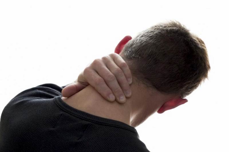
Did you wake up this morning with acute facial pain and inability to open your mouth? When you think to yesterday, you remember taking a big bite from an apple and experienced a sharp pain in your jaw joint. Today, you can’t really open or fully close your mouth. That unexplained clicking in your jaw that you’ve had for two years has suddenly gone away. Now, there’s just facial tenderness in front of your ear and you’re worried that you can’t eat! If you experience this, you likely have a “locked jaw”, or Acute Disc Displacement. This is one of the common disorders of the Temporomandibular Joint, more known as the TMJ.

The TMJ is the ball and socket joint that connects the Mandible (jaw bone) and the Temporal bone (one of the bones of your skull). It’s the small joint located in front of your ear. There is a cartilage cushion in between the ball and socket, referred to as the Disc. The disc is supported by special Ligaments, which keep the disc in place. Movement problems of the disc can be responsible for creating many symptoms in the TMJ, such as clicking, crepitations, locking, muscle spasm, and pain.

TMJ Disorder/Dysfunction, or TMJD/TMD, is seen more commonly in women than men. There is a 3:1 incidence in females to males, and can include one or both jaw joints. In most instances, the dysfunction is a result of an imbalance or change in the normal function of the bones, ligaments, muscles, disc, or nerve components of the TMJ complex.

Disc Displacement is a mechanical problem that occurs when the disc ends up in the wrong position within the ball and socket. In this case, it is likely that the disc has become displaced due to the wide opening, creating increased stress and strain on the ligaments, resulting in pain around the joint and spasm in the facial muscles. There is also a longstanding history of unexplained clicking in the joint, which may be a pre-disposing factor to this problem. In essence, the disc needs to be properly re-educated to find its’ normal resting position again, and the mechanics restored to the muscles and joint. Specialized physical therapy care would assist in restoration of disc position, to restore movement and function, as well as normalize the pain.

Dysfunction in these tiny joints can have many causes. These can include trauma such as blow to the head, whiplash, falls, sports injuries, arthritis, dental occlusion, dentures, history of joint noises, stress (clenching, grinding), poor habits (posture, physical, oral), and many others. Signs and symptoms may include facial pain, headache, neck pain, ear pain, sudden changes in bite, tooth pain, ringing in the ears, shoulder pain, and facial tingling or numbness.

A physical therapist can help you understand and manage your TMJ dysfunction. Often it is obvious that the TMJ is the cause of symptoms you experience. However, for many people, the symptoms they experience can be widespread, and may not make it so obvious. As mechanics for the human body, physical therapists can properly assess, diagnose, and treat the TMJ and most of the possible influences. We provide a specialized hands-on approach to treatment, education, and exercise program. Treatment may also include other muscle re-education techniques such as Intramuscular Stimulation (IMS). In many instances, we often work with your oral practitioner (dentist, orthodontist, oral surgeon) to ensure the best outcome possible for your TMJ. Treatment can be very effective in a short period of time. A self-applied approach with a little guidance from specialized physical therapy practitioners will help with long-term benefits.

Acute Disc Displacement is only one of several diagnoses of TMJ dysfunction. Keep connected to read about other problems that these tiny joints can give you. Don’t stop eating apples though…

Vince Cunanan is a registered physiotherapist and associate at Sun City Physiotherapy’s Downtown Kelowna and Glenmore clinics. He can be contacted at info@suncityphysiotherapy.com

Bone Health and Exercise

Bone Health and Exercise

Physiotherapy » Posts Tagged "glenmore physiotherapy" (Page 4)

Bone Health and Exercise

Optimal bone health is a serious consideration in people approaching mid-life. It is common knowledge that bones become more brittle as we age. What may be less recognized are the factors we can control through exercise and nutrition in the first half of adult life that have a direct effect on prevention of osteoporosis.

Osteoporosis is a disease characterized with a loss in bone mass and deterioration in the sturdiness of bone structure. This is associated with the increased risk of fractures, particularly in the hip, wrist and spine. It is prevalent in the older population. If you are a woman, you have a 50% lifetime risk of a fracture from osteoporosis.
Bone loss is an insidious process. In the early stages it is called “osteopenia” It can occur when an arm or leg is casted after a frracture. A loss in bone mineral density (BMD) can accelerated after taking steroid medications or with certain autoimmune disorders or diabetes.

Reduced levels of estrogen after menopause accelerate bone loss. However, it is well- established that if a woman in her 40’s and 50’s exercises regularly and has good nutritional habits, she can diminish her post-menopausal bone density loss substantially. Osteoporosis is optional!

How does exercise affect bone health?
Bone is a living tissue. Bone cells, called osteophytes, have the ability to act like strain gauges and adapt to the amount of stress placed on them. Regular weight bearing exercise or strength training is essential to maintaining healthy bone. Resistance training improves muscle mass and strength and can increase spine and hip bone density. It is essential that the exercises chosen are safe and appropriate for the individual. Physiotherapists assess posture, understand risk factors and can advise which exercises are best for those with osteoporosis.

As a preventative measure and for those with mild osteopenia, high and medium impact exercise such as soccer, tennis, activities such as skipping and step-ups can stimulate healthy bone cells to produce a stronger bony matrix and increase BMD.

For individuals with moderate osteoporosis, weight bearing exercise and moderate impact exercises are appropriate, high impact exercise is not. Strength training exercises should target the specific areas affected. Balance exercises and fall prevention awareness are important, as falls can result in fractures, which are painful and can take much longer to heal than with normal bone. Working with a physiotherapist with specific knowledge about osteoporosis to set up an exercise program is highly recommended. Check with your physician before beginning a strenuous exercise program.

Attention to postural alignment during strength training is important. Certain exercises place too much strain on the midback area and can increase the rounding, or kyphosis in this area. For example, repeated curl-ups for abdominal strengthening, or swinging kettle balls with arms extended should be avoided.

Why does nutrition play such an important role in prevention of bone mineral density loss?
Bones are important warehouses for calcium and other important minerals needed for cellular function. If our diet is low in calcium, the body borrows it from our bones. If it’s not restored, a net deficit in minerals can result in reduced BMD.
In order to maintain optimal bone health, sufficient amounts of Calcium, Magnesium, Vitamin D are recommended.

Sun City Physiotherapy Locations

Downtown

1468 St. Paul Street, Kelowna, BC
Phone: 250-861-8056
downtown@suncityphysiotherapy.com
more info

Glenmore

103-437 Glenmore Road, Kelowna, BC
Phone: 250-762-6313
glenmore@suncityphysiotherapy.com
more info

Lake Country

40-9522 Main St., Lake Country, BC
Phone: 250-766-2544
winfield@suncityphysiotherapy.com
more info

Lower Mission

3970 Lakeshore Road, Kelowna, BC
Phone: 778-699-2006
lowermission@suncityphysiotherapy.com
more info
Classification of Whiplash Injuries

Classification of Whiplash Injuries

Physiotherapy » Posts Tagged "glenmore physiotherapy" (Page 4)

Classification of Whiplash Injuries

Motor vehicle accidents (MVA) are sometimes an unfortunate consequence of modern reliance we have on automobile transportation. Previous to the invention of the car, there was “railroad neck”, and also a whiplash variation termed “roller-coaster neck”. Motor Vehicle accidents are sometimes termed “motor vehicle crashes” to suggest the preventable nature of some accidents. When a crash does occur, there is usually a chain of events including visits to the auto body shops, ongoing communication with insurance claims agents, and visits to many different health care professionals.

Despite advancement in sophisticated safety equipment for cars, bodily injury can sometimes result from accidents. Although the impact can occur in less than a second, the resulting injuries can persist for weeks or months. The neck (cervical spine) is the most common area of involvement, however, the mid back or low back can also sustain trauma. The shoulder or chest area can also be bruised by the restraint effect of the seat belt.

Injuries to the cervical spine are termed “whiplash-associated disorder’ or WAD for short. As with most conditions and injuries, there is a spectrum of severity. Following much consultation, the Quebec Task Force on Clinical Classification of WAD developed a five point scale of severity in 1995, which is well recognized within the health care and insurance fields. With Grade 0, or WAD 0, there are no physical signs, and the person does not complain of symptoms or usually seek help. At the other extreme end of the spectrum is the most severe WAD IV injury in which X-rays indicate a fracture or dislocation and require urgent medical attention. WAD classification is determined by a detailed and comprehensive clinical examination. Fortunately, most of the whiplash injuries sustained in a crash are classified as WAD II or III, and are commonly treated in physiotherapy practice. Clinical features include pain, and limited range of motion due to muscle spasm, sprained ligaments, and inflamed joints. Irritation of the neurological system can manifest as symptoms of numbness or tingling extending into the upper or lower extremities.

Physical therapists are very familiar with performing a detailed assessment and can help with determining the degree of injury that a person has sustained. A physiotherapy treatment plan is determined based on the degree of injury and time since the accident. Early in treatment when the condition is still very acute, it is common to suggest use of ice to decrease inflammation and pain relieving physical electrical modalities can also be used. As the person’s symptoms decrease, gentle mobilization, range of motion exercises, and progressive strengthening exercises are commonly introduced. Progress with treatment is individual and dependent on many factors. There are numerous practice guidelines, however, which suggest a return to normal daily activities as soon as possible, and treatment that promotes return of function is most helpful. A physical therapist is well trained to help assess your whiplash condition and provide an individualized treatment plan.


Sun City Physiotherapy Locations

Downtown

1468 St. Paul Street, Kelowna, BC
Phone: 250-861-8056
downtown@suncityphysiotherapy.com
more info

Glenmore

103-437 Glenmore Road, Kelowna, BC
Phone: 250-762-6313
glenmore@suncityphysiotherapy.com
more info

Lake Country

40-9522 Main St., Lake Country, BC
Phone: 250-766-2544
winfield@suncityphysiotherapy.com
more info

Lower Mission

3970 Lakeshore Road, Kelowna, BC
Phone: 778-699-2006
lowermission@suncityphysiotherapy.com
more info

Carpal Tunnel Syndrome

Carpal tunnel syndrome is a common wrist and hand injury.  It occurs when one of the major nerves called the median nerve is compressed within the carpal tunnel of the wrist.  This may occur for a number of reasons but some of the more common reasons include: pregnancy, gout, trauma (repetitive or direct trauma), or infection.  Carpal tunnel can happen at anytime, however it is more common between the ages of 40-60 and women tend to be affected more often then men.

This condition is characterized by burning wrist pain and numbness or tingling within the hand.  This often occurs at night and the patient usually wakes up due to the symptoms.   In some cases the pain can radiate to the forearm, elbow, and shoulder.

Some of the other symptoms reported by patients include poor sensation in the hand, weakness of the hand, cramping in the hand, reduced temperature in the hand, and sometimes shaking or flicking the hand relieves the symptoms.  There may also be muscle wasting around the thumb.  Movements of the hand are often pain free, however some resisted movements around the thumb can be painful.

Diagnosing carpal tunnel syndrome is often done based on the patient’s medical history.  Your physiotherapist or physician will take you through a series of tests and if carpal tunnel syndrome is suspected a nerve conduction test may be ordered to confirm the diagnosis.  Diabetes should be excluded, as it can be a risk factor for carpal tunnel syndrome.

Mild cases can be treated conservatively.  This may include splinting, activity modification, diuretics, and NSAID medications.  Techniques such as contrast baths, which are alternating baths of cold and warm water, may be used in some cases to control inflammation and swelling.  A physiotherapist may give you specific exercises that can be helpful with a patient’s recovery.  For more persistent or severe cases, corticosteroid injections or surgery may be required.

Wrist and hand injuries are often difficult to diagnose so if you are experiencing symptoms of this nature it is recommended that you see your health care professional to have it taken care of.